case of the
month
2019
COTM DECEMBER 2019
Case from Dr Amandeep Mann and Mr Keith Jones (Royal Derby Hospital, UK)
CLINICAL DETAILS
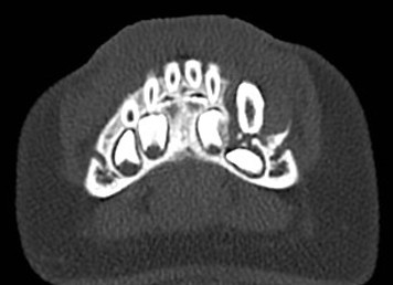
A 2 year old girl presented with a mass invlving the left mandible. A CT scan showed an expansile lesion involving the left mandible parasymphysis closely associated with and encapsualting primary and permanent teeth. There is resorption and displacement of adjacent structures
(see images below).


The lesion was biopsied and sent for histopathological examination (click on the link below).
Scroll to the right for details and diagnosis.
COTM NOVEMBER 2019
Case from Dr F Tahir (Royal Hallamshire Hospital, Sheffield, UK)
CLINICAL DETAILS
A 33 year old fit and well male patient presented with a largely painless swellinginvolving the left upper lip. An MRI scan of the head and neck region showed a well defined mass centred on the left nasolabial fold with pressure resorption of bone focally (see image below).

The lesion was enucleated and sent for histopathological examination (click on the link below).
Scroll to the right for details and diagnosis.
COTM OCTOBER 2019
Case and Images from Dr A Levene and Mr CH Chan (Spire Bushey Hospital, UK)
CLINICAL DETAILS
A 46 year old female presented with a 1 cm lump involving the gingivae in the LR3 region. The patient was fit and well. The lesion was firm, asymptomatic and not related to gum disease (see images below).


The cone beam CT shows a well-defined and corticated mixed density lesion in the LR3-4 region with a central intraosseus component and expansion resorption of the buccal cortex. An incisional biopsy of the lesion was sent for histopathological examination (click on the link below).
Scroll to the right for details and diagnosis.
COTM AUGUST/SEPTEMBER 2019
CLINICAL DETAILS
A 60 year old male presented with a history of a recurrent swelling involving the right parasymphysis of mandible in the LR5 region. The patient was fit and well. The LR5 was extracted a few months back however, the symptoms and swelling at the site had not resolved (see image below).

The panoramic radiograph shows a well-defined radiolucent lesion in the LR5 region lacking corticationwith resorption of the overlying mandibular cortex. An incisional biopsy of the lesion was sent for histopathological examination (click on the link below). Scroll to the right for details and diagnosis.
COTM JULY 2019
CLINICAL DETAILS
A 50 year old female with a root filled UR1 presented with a painless midline palatal swelling. The patient was diabetic and on metformin. Cone beam computed tomography (CBCT) of the anterior maxilla showed a round unilocular radiolucency in the midline. The lesion was well-defined and well-corticated and extended across both sides of the incisive canals (see image below).

An incisional biopsy of the lesion was sent for histopathological examination (click on the link below).
Scroll to the right for details and diagnosis.
Virtual Microscopy/Whole Slide Image (H&E)
COTM JUNE 2019
CLINICAL DETAILS
A 66 year old male patient presented with a six week history of a white lesion involving the right lateral border of tongue. The lesion was largely asymptomatic and had a somewhat irregular appearance. The patient smoked at least 10 cigarettes per day.

An incisional biopsy of the white patch was sent for histopathological examination
(click on the link below). Scroll to the right for details and diagnosis.
COTM MAY 2019
CLINICAL DETAILS
A 44 year old male patient presented with a painless cyst in the lower right mandible(LR7/8 region).

The lesion was enucleated and the two extracted molar extracted. Tissue was sent for histopathological examination (click on the link below). Scroll to the right for details and diagnosis.
COTM APRIL 2019
CLINICAL DETAILS
A 54 year old black woman presented with firm bony jaw swelling in both the upper right and lower left quadrants. The radiological images can be seen below.


The patient underwent an excisional biopsy of the tissue in the upper right quadrant
(click on link below). Scroll to the right for details and diagnosis.
Virtual Microscopy/Whole Slide Image 1
COTM MARCH 2019
CLINICAL DETAILS
An 82 year old male patient presented with a three week history of multiple ulcers involving the hard palate, gingivae and floor of mouth. The patient had a history of a liver transplant in 1987 and hypertension.


The ulcers were biopsied and sent for histopathological examination (click on the link below).
Scroll to the right for details and diagnosis.
Virtual Microscopy/Whole Slide Image (H&E)
COTM FEBRUARY 2019
CLINICAL DETAILS
A 29 year old female presented with an approximately 2 cm swelling in the right floor of mouth which appeared submucosal and well defined. The patient was fit and well.


The lesion was excised and sent for histopathological examination (click on the link below).
Scroll to the right for details and diagnosis.
Virtual Microscopy/Whole Slide Image (H&E)
COTM JANUARY 2019
CLINICAL DETAILS
A 7 year old boy presented with a history of discoloured promary teeth. There was a family history of problem affecting teeth. The erupted permanent teeth also showed severe hypomineralisation and pitting. The patient was fit and well with no childhood caries or vitamin deficiencies.


The exfoliated teeth were sent for histopathological examination (click on links below).
Scroll to the right for details and diagnosis.
Virtual Microscopy/Whole Slide Image (Ground Section)
